“Cancer cells have a range of tools at their disposal to resist the effects of powerful chemotherapy drugs,” said Evripidis Gavathiotis, PhD, Co-Leader, MECC Cancer Therapeutics Program, Professor, Biochemistry and Medicine, Albert Einstein College of Medicine, and co-corresponding author of the paper. “Based on our extensive study of apoptosis, also known as programmed cell death, we’ve developed a new strategy for making existing treatments against AML more effective.”
Killing Cells by Puncturing Mitochondria
Chemotherapy and radiation therapy both work by damaging cancer cells severely enough to make them undergo apoptosis. When cancers become resistant to therapy, impaired apoptosis is almost always the reason.
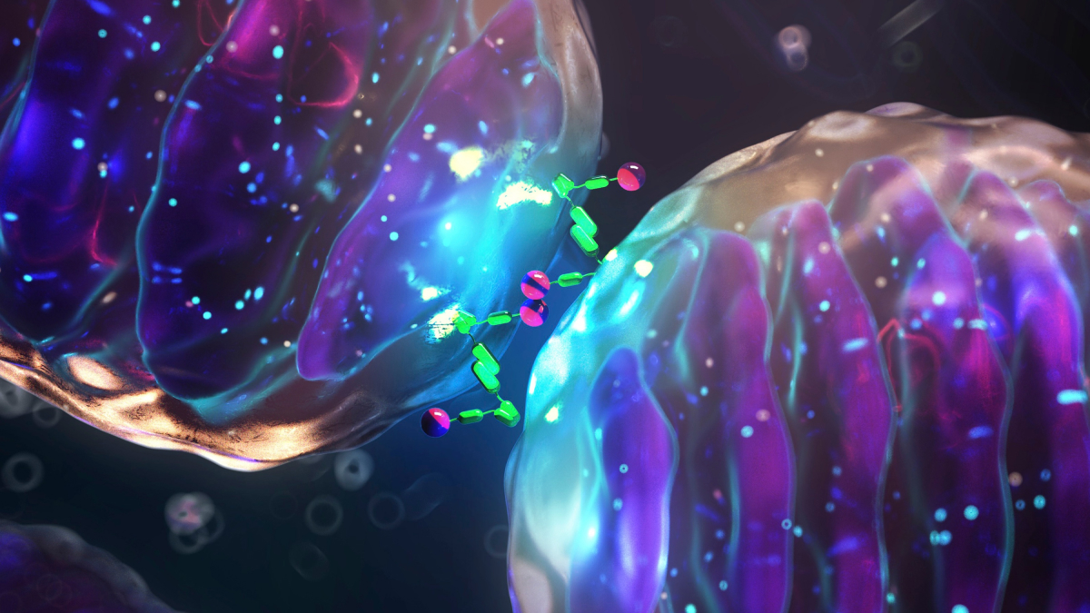
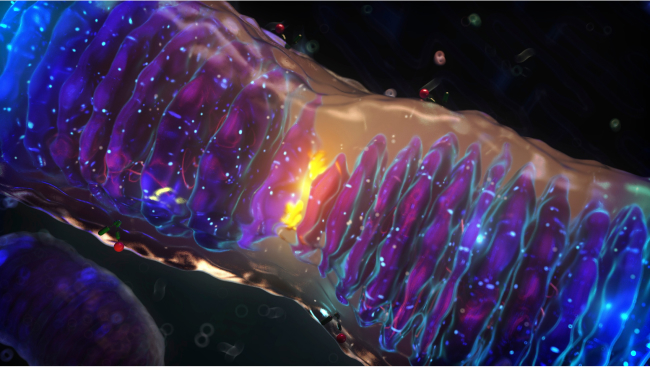
The body relies on apoptosis to get rid of unwanted cells—excess cells pruned during embryological development, for example. Apoptosis is a highly choreographed sequence of molecular events that requires damage to mitochondria, the critical intracellular components that produce the energy that enables cells to function. Each cell contains hundreds of mitochondria, and their elimination—caused by a protein that punctures them—is the key step in mediating cell death via apoptosis.
For optimal functioning, mitochondria depend on their ability to fuse with each other—something they do when they need to produce more energy or to function under stressful conditions. Fusion is regulated by two specialized proteins, called mitofusins, which reside on the outer membrane of mitochondria. Health problems can occur when mitofusins are defective. “We had no way to alter mitofusin activity, making it difficult to understand how they influence mitochondria,” said Dr. Gavathiotis.
A Focus on Fusion
With his expertise in structural and chemical biology, Dr. Gavathiotis designs small molecules to serve as “tools” for biological research or as prototypes for new drugs. In a study published last July in Nature Communications, his research team synthesized small molecules that enabled them to either activate or inhibit mitofusins. Their mitofusin activator stimulated mitochondria to fuse and improved their function; in contrast, their mitofusin inhibitor prevented mitochondria from fusing with each other and impaired their function.
“The results we observed with our mitofusin inhibitor, MFI8, were especially intriguing,” Dr. Gavathiotis recalled. “MFI8 not only prevented mitochondrial fusion but also—by fragmenting cells’ mitochondria—primed cells to undergo apoptosis.”
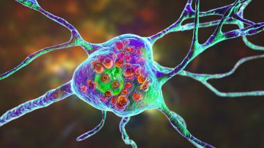
Dr. Gavathiotis reasoned that MFI8 might be able to sensitize cancers to the apoptosis-promoting chemotherapies to which they had become resistant. He decided to test his hypothesis on AML, which has a dismal five-year overall survival rate of only 28%.
Finding Why Resistance Occurs
In 2018, the U.S. Food and Drug Administration (FDA) approved a promising two-drug therapy for treating AML. While early results were promising, prolonged treatment with the drug combination has led to drug resistance.
“To determine whether our mitofusin inhibitor MFI8 could help in treating AML, we first had to discover how AML becomes resistant to therapy, including to the recent FDA-approved treatment,” Dr. Gavathiotis said. His colleagues found that resistance could be explained, in part, by a quality-control system that cells use to keep their mitochondria healthy.
When mitochondria undergo extreme stress (from exposure to chemotherapy, for example), the stress activates a process called mitochondrial autophagy, or mitophagy, that targets and digests damaged mitochondria.
The researchers found that mitophagy in drug-resistant human AML cells happened at a higher rate than in drug-sensitive AML cells. “With their increased rate of mitophagy, the drug-resistant AML cells were able to maintain a healthy and functional population of mitochondria,” Dr. Gavathiotis said. “In those cancer cells, chemotherapy wasn’t able to cause enough mitochondrial damage to induce the cells to undergo apoptosis and die.”